What is a CT (Computed Tomography) Scan?
Category: Procedures, Spine | Author: Stefano Sinicropi
 Note: this is a companion article to yesterday’s post on What is an MRI?
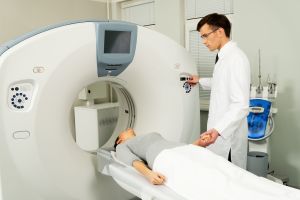
A CT scan is a method of medical imaging that is used to assess bone structure and soft tissue issues. X-rays are used to collect the image. You may have heard of this referred to as a CAT (Computed Axial Tomography) scan in the past.
To obtain a CT scan, a radiology technician will have you lie on a table so you can be passes through the scanner. The x-ray source rotates around you, and the scanner saves the data and generates the image that results. This called a “helical” scan, and it creates images that can be viewed as “slices” of the area being scanned. CT scans are usually brief, and may or may not require radiological contrast. Please advise the technician if you have ever had a reaction to any medical contrasts.
What does the CT Scan Tell my Provider?
In the context of spine care, a CT scan can be used to evaluate a number of things:
- How well-healed a spinal fusion is
- Whether or not compression fractures are present (these can be difficult to see on normal x-rays)
- To image soft tissues such as intervertebral discs in patients who cannot have MRI scans due to medical devices or implants
Will the CT Scan Expose me to Large Amounts of Radiation?
Yes. X-rays are a form of ionizing radiation, and this exposure is a risk with CT scans. That said, most modern CT scanners are designed to expose patients to less radiation than in the past.
CT Scan Risks vs. Benefits
Here’s a link to the National Cancer Institute’s CT scan information page. It has more in-depth information that’s beyond the scope of this blog post.