What is an MRI?
Category: Minimally Invasive Surgery, Procedures | Author: Stefano Sinicropi
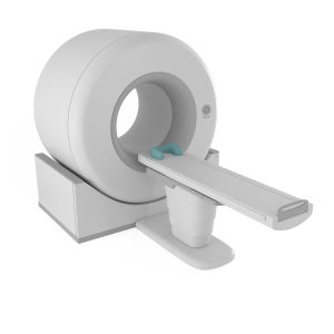
 Magnetic Resonance Imaging (MRI) is a method of medical imaging. It allows health care providers to assess soft tissues and bones without an invasive procedure.
MRI machines use a powerful magnet and radio-frequency sound waves to produce the images ordered by your provider. The magnet and radio waves interact with water and other molecules in your body that move in response. The MRI detects these interactions and creates the image via software.
In the context of spine imaging, MRI scans are most frequently used to assess potential sources of pain, weakness, numbness, or tingling. This includes, but isn’t limited to:
- Whether or not the spinal cord is being pressed, or impinged, by tissue or bone changes
- Whether or not the nerves exiting the spine are being compressed or “pinched”
- How severely any compressed nerves are being pinched
- If a disc is herniated or bulging in such a way as to compress nerves
- If there are inflamed, arthritic areas present in the vertebral joints
For some imaging, your provider may request that contrast be used. Contrast will emphasize fluid flows in the area of interest. This is more commonly used outside spinal areas.
How can I Find out the Results of my MRI?
Usually, the provider who ordered the MRI, or one of their colleagues, will review the image on their own, along with a reading report provided by a radiologist. Then, the provider will go over the results with you. Some providers do this in clinic, and others do it by phone.
How can I Request a Copy of my Imaging?
Most imaging centers will provide you with a CD of your images on request.