What Is A Spinal Myelography?
Category: Spine | Author: Stefano Sinicropi | Date: May 2, 2019
When it comes to making an accurate diagnosis of your back condition, spine experts have a number of tools in their arsenal to help them make an accurate conclusion. Imaging techniques allow them to see more than what’s possible with the naked eye, and one such imaging technique is called a spinal myelography. Below, we take a closer look at the technique and explore its benefits and risks.
Spinal Myelography Basics
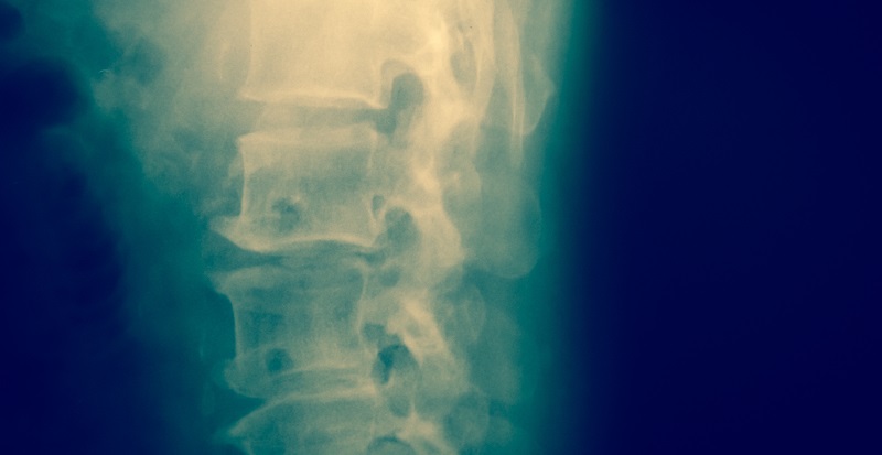
Myelography involves a real-time form of x-ray called a fluoroscopy and a contrast injection that allows the surgeon to view structures like the spinal cord, the spinal nerve roots or the meninges. It also can be used after a surgery has been performed to ensure everything in the spinal system is working as expected.
During the procedure, a needle full of contrast dye will be inserted into the spinal canal. The dye allows the surgeon to use x-ray technology to view structures that wouldn’t be visible on a standard x-ray exam. The doctor will take x-ray images and use a fluoroscope to view real-time images of the spinal cord and surrounding nerves. This allows them to get a comprehensive understanding of exactly what’s causing your discomfort.
Spinal myelography is often used to look for and diagnose the presence of:
- Disc herniation
- Nerve root compression
- Nerve or disc degeneration
- Tumors
- Infections
- Inflammation
Risks and Benefits
Like most diagnostic techniques, a spinal myelography offers some unique benefits and carries some risks. For example, one reason why spinal myelography is often ordered is because it can be used in situations where the insertion of spinal hardware makes it such that an MRI cannot be used. The myelography allows the doctor to view previously inserted hardware to ensure it’s in the correct location and working as it should. Because an MRI uses magnetic waves to produce an image, it can’t be used if a person has certain metal hardware in their body.
Other benefits of a spinal myelography include:
- It’s a relatively safe and painless procedure.
- Allows viewing of areas of the spine that are not distinguishable on other image techniques.
- No radiation remains in the patient’s body after the x-ray.
- Usually no side effects of radiation when used in the diagnostic range for myelography.
As with any procedure, there are also some potential risks and side effects. Some people experience headaches or nausea following the contrast injection, but this is rare. Other potential risks include:
- Very rare chance of problems caused by radiation exposure, like cancer. However, the benefits far outweigh the potential risk.
- Failed clotting of the epidural space can lead to headaches when standing. The puncture site usually heals on its own, but if the opening remains and spinal fluid leaks out of the spinal canal, headaches may develop. This can be addressed with an epidural patch if necessary.
- Potential adverse reaction to the contrast dye.
- Nerve injury or bleeding.
These risks are very rare, and the benefits of the procedure almost always outweigh any potential complications. Dr. Sinicropi would be happy to walk you through anything else related to the procedure or answer any questions you might have by contacting his office for more information.